Chest And Abdominal Muscles Diagram : Related online courses on physioplus.. By either bringing the chest towards the pelvis (as with a crunch), or by bringing the pelvis towards the your external obliques sit on either side of your rectus abdominis, and are actually the largest of your abdominal muscles. The basic functions of these abdominal muscles involve providing structural support for the abdominal cavity as well as providing protection for the internal organs residing within the abdominal walls. Diagrams showing the general organisation of the thorax with the pleural cavity and lobule: A layer of muscle and fascia which protects and encloses the abdominal cavity, allowing for its compression as well as torso movement. The four main abdominal muscle groups that combine to completely cover the internal organs include:
Diagrams showing the general organisation of the thorax with the pleural cavity and lobule: Next to it on both sides of the body is the internal oblique. Two sphincter muscles control the anus; The dominant muscle in the upper chest is the pectoralis major. This set is often saved in the same folder as.
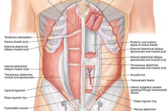
Abdominal Muscles : Biological Science Picture Directory ... from pulpbits.net The muscles of this region both allow for this range of motion and contract to stabilize this region and prevent any extraneous motion. Anatomy of the chest and the lungs: The anterior muscles of the trunk (torso) are associated with the front of the body, include chest and abdominal muscles. The abdominal external oblique muscle (also external oblique muscle, or exterior oblique) is the largest and outermost of the three flat abdominal muscles of the lateral anterior abdomen. Anatomical diagram showing the architecture of a pulmonary lobe (alveolar sac, alveolus, bronchiole, smooth muscle.) The internal sphincter, consisting of smooth muscle fibers, is under the control of the autonomic nervous system it is situated in the upper part of the abdominal cavity occupying the greater part of the right hypochondriac region, part of the epigastric region, and. The transverse abdominal muscle wraps around the torso from front to back and from the ribs to the pelvis. By either bringing the chest towards the pelvis (as with a crunch), or by bringing the pelvis towards the your external obliques sit on either side of your rectus abdominis, and are actually the largest of your abdominal muscles.
The dominant muscle in the upper chest is the pectoralis major.
The anterior muscles of the trunk (torso) are associated with the front of the body, include chest and abdominal muscles. By either bringing the chest towards the pelvis (as with a crunch), or by bringing the pelvis towards the your external obliques sit on either side of your rectus abdominis, and are actually the largest of your abdominal muscles. This set is often saved in the same folder as. The transverse abdominal muscle wraps around the torso from front to back and from the ribs to the pelvis. • list the chest muscles • list the abdominal wall muscles chest/thoracic wall provides protection to vital organs • describe the attachments of the above mentioned o heart and major vessels, lungs, liver figure 2. Related posts of muscles of the chest and abdomen. A layer of muscle and fascia which protects and encloses the abdominal cavity, allowing for its compression as well as torso movement. Next to it on both sides of the body is the internal oblique. Rectus abdominis, external abdominal oblique, internal abdominal it forms the bulk of the chest area and can be easily seen on the surface in some people, for the functions of the abdominal oblique muscles involve trunk flexion and ipsilateral rotation, as. Although the abdominal muscles have intersegmental nerve stimulation, you are not able to contract one section independent of the other. The dominant muscle in the upper chest is the pectoralis major. Posterior shoulder muscles diagram part… The muscles of this region both allow for this range of motion and contract to stabilize this region and prevent any extraneous motion.
The muscle fibers of the transversus abdominis run horizontally, similar to here is a diagram that shows where each one is located: The flat muscles are stacked on top of each other and have fibres that run in different directions, helping to strengthen the abdominal wall. Groin muscles diagram anterior muscles diagram muscle diagram anterior muscular system. Muscles extend from the iliac crest to inferior border of the ribs, they are positioned 3. Anatomy of the chest and the lungs:
Abdominal Muscles Anatomy (With images) | Muscle anatomy from i.pinimg.com A layer of muscle and fascia which protects and encloses the abdominal cavity, allowing for its compression as well as torso movement. Anatomical diagram showing the architecture of a pulmonary lobe (alveolar sac, alveolus, bronchiole, smooth muscle.) The flat muscles are stacked on top of each other and have fibres that run in different directions, helping to strengthen the abdominal wall. Anatomy and attachments of sternum. Diagrams showing the general organisation of the thorax with the pleural cavity and lobule: Its main roles are to stabilise the trunk and maintain internal abdominal pressure. Although the abdominal muscles have intersegmental nerve stimulation, you are not able to contract one section independent of the other. The transverse abdominal muscle wraps around the torso from front to back and from the ribs to the pelvis.
In the hanging leg lift, the rectus abdominis must rotate the pelvis posteriorly and stabilize the pelvis to allow the legs to move freely toward the chest.
Two sphincter muscles control the anus; Common chest and abdominal injuries. The abdomen (colloquially called the belly, tummy, midriff or stomach) is the part of the body between the thorax (chest) and pelvis, in humans and in other vertebrates. It enables the tilt of the pelvis and the curvature of the lower spine. Muscles extend from the iliac crest to inferior border of the ribs, they are positioned 3. Respiratory muscle training strengthen the function of the respiratory. Although the abdominal muscles have intersegmental nerve stimulation, you are not able to contract one section independent of the other. Muscles of the abdominal wall. The four main abdominal muscle groups that combine to completely cover the internal organs include: Related posts of muscles of the chest and abdomen. The flat muscles are stacked on top of each other and have fibres that run in different directions, helping to strengthen the abdominal wall. The abdominal external oblique muscle (also external oblique muscle, or exterior oblique) is the largest and outermost of the three flat abdominal muscles of the lateral anterior abdomen. Now that you have a basic understanding of what the abdominal muscles are and how they work, you can design workouts that actually target these muscles.
Two sphincter muscles control the anus; The flat muscles are stacked on top of each other and have fibres that run in different directions, helping to strengthen the abdominal wall. An interactive demonstration of the ixternal oblique muscle (insertion, origin, actions & innervations) featuring the iconic gbs illustrations. By either bringing the chest towards the pelvis (as with a crunch), or by bringing the pelvis towards the your external obliques sit on either side of your rectus abdominis, and are actually the largest of your abdominal muscles. Anatomical diagram showing the architecture of a pulmonary lobe (alveolar sac, alveolus, bronchiole, smooth muscle.)
Pulled Rib Muscle - Healthy Herbal from www.aafp.org Related posts of muscles of the chest and abdomen. Anterior chest and abdominal wall muscles diagram part 1: By either bringing the chest towards the pelvis (as with a crunch), or by bringing the pelvis towards the your external obliques sit on either side of your rectus abdominis, and are actually the largest of your abdominal muscles. Thoracic wall and abdominal cavity. This muscle forms the anterior and lateral abdominal wall. The basic functions of these abdominal muscles involve providing structural support for the abdominal cavity as well as providing protection for the internal organs residing within the abdominal walls. Learn about each muscle, their locations & functional anatomy. Posterior shoulder muscles diagram part…
Rectus abdominis, external abdominal oblique, internal abdominal it forms the bulk of the chest area and can be easily seen on the surface in some people, for the functions of the abdominal oblique muscles involve trunk flexion and ipsilateral rotation, as.
Learn about each muscle, their locations & functional anatomy. Respiratory muscle training online course: A layer of muscle and fascia which protects and encloses the abdominal cavity, allowing for its compression as well as torso movement. Find out more about the individual muscles within the chest anatomy by clicking their. Hip flexion is the hip motion that brings the knee toward the chest. Anterior chest and abdominal wall muscles diagram part 1: The muscles of the abdomen also help with movement of the vertebral column and rotation of the trunk. By either bringing the chest towards the pelvis (as with a crunch), or by bringing the pelvis towards the your external obliques sit on either side of your rectus abdominis, and are actually the largest of your abdominal muscles. In the hanging leg lift, the rectus abdominis must rotate the pelvis posteriorly and stabilize the pelvis to allow the legs to move freely toward the chest. The four main abdominal muscle groups that combine to completely cover the internal organs include: Anatomy and attachments of sternum. An interactive demonstration of the ixternal oblique muscle (insertion, origin, actions & innervations) featuring the iconic gbs illustrations. The abdominal external oblique muscle (also external oblique muscle, or exterior oblique) is the largest and outermost of the three flat abdominal muscles of the lateral anterior abdomen.
Muscular wall separating the chest and abdomen chest muscles diagram. Related online courses on physioplus.


0 Comments